


 Back
BackIt’s Flu Season, even for horses!
Equine influenza is a highly contagious respiratory disease that continues to cause outbreaks in equids worldwide, despite widespread vaccination.
Rapid and accurate diagnosis of equine influenza is essential to limit virus spread, protect animal health, and ensure compliance with movement and competition requirements.
Today, effective equine influenza control relies on a combination of PCR testing and serological surveillance. Real-time PCR enables early detection of the virus, even before clinical signs appear or in vaccinated horses while serology can be useful for retrospective investigations (demonstrating seroconversion between paired samples) and epidemiological studies.
By combining molecular diagnostics and serology, veterinarians and laboratories can strengthen surveillance, support informed vaccination and biosecurity decisions throughout the equine industry. Differentiation between infected and vaccinated horse is possible when the appropriate vaccine is used.

1. What Is Equine Influenza and Why Surveillance Matters
a) The Causative Agent:
Equine influenza is caused by the Equine Influenza Virus (EIV). Two EIV subtypes have historically infected horses: H7N7 and H3N8. While the H7N7 has not been reported since 1980, H3N8 subtypes still have major sanitary and economic impacts on the equine industry worldwide. Currently only Florida sublineage clades 1 and 2 are circulating in Europe. Clade 1 commonly circulates in North America (see figure 1).
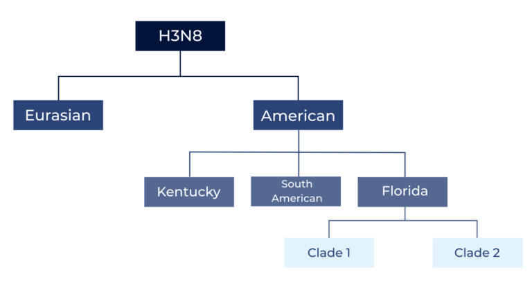
Figure 1: EIV H3N8 split into American and European lineages; the American lineage further diversified into Kentucky, South American, and the predominant Florida sublineage, which later divided into Florida clades 1 and 2.
b) Transmission and symptoms:
Equine influenza is extremely contagious and can spread rapidly within a horse population. Transmission occurs mainly through direct horse-to-horse contact, although indirect spread via contaminated equipment or people is also possible, despite the virus having limited survival in the environment.
After an incubation period of 2–5 days, affected horses typically develop a high fever (often above 39.5 °C), depression, reduced appetite, and respiratory signs, including a harsh dry cough and a clear, watery nasal discharge, which turns thick and green in colour after four to five days.
c) Control of the disease
Control of equine influenza depends on early detection, strict biosecurity, and preventive strategies. Vaccination plays a key role to limit outbreaks and reduce disease severity, with booster doses generally recommended every 6–12 months depending on risk exposure and regulatory requirements. However, vaccinated horses that become infected may still shed the virus and contribute to transmission within the group.
2. Laboratory Diagnosis of Equine Influenza: Pathogen detection and serology
Laboratory diagnostics are essential to confirm infection, monitor outbreaks, and support surveillance programs. Common diagnostic approaches include:
- Pathogen detection by real-time PCR (RT qPCR): the most sensitive and rapid method for detecting viral RNA. RT qPCR allows for early identification of infected animals, even before clinical signs appear and under vaccinal protection. It is used to demonstrate freedom from infection prior to movement, sale or competition.
- Virus isolation – Performed in embryonated chicken eggs or cell culture. While confirmatory, this method is time-consuming and less suitable for rapid decision-making.
- Antigen detection assays – Including ELISA or rapid lateral flow tests, useful for field screening though less sensitive than RT-qPCR.
- Serology – Detection of antibodies using Hemagglutination Inhibition (HI), Single Radial Hemolysis (SRH), or ELISA. These methods are particularly valuable for retrospective diagnosis and epidemiological surveillance.
Combining molecular and serological tools helps veterinarians to assess infection status, guide vaccination strategies, and reduce the risk of disease spread in stables, breeding farms, and competition settings.
3. Supporting Equine Influenza Surveillance with IDvet Diagnostic Solutions
a) Monitor the presence of Influenza A virus using real time PCR, the most sensitive and rapid method for detecting viral RNA
The ID Gene Lyo™ Influenza A Multi-species Triplex qPCR allows the simultaneous detection of EIV along with two non-target positive controls (endogenous and exogenous), ensuring result reliability.
The assay is validated for use on equine nasal or nasopharyngeal swabs and supports fast, confident decision-making.
b) Serology: Monitor exposure and vaccination compliance
ELISA-based antibody detection is a simple and effective method to assess seroprevalence at herd or population level. Using the highly conserved Influenza A nucleoprotein (NP), the ID Screen® Influenza A Antibody Competition ELISA can detect anti-NP antibodies in serum or plasma from multiple species, including equids. It delivers faster results than traditional serological tests (HI, SRH) and identifies both IgM and IgG antibodies, enabling early as well as long-term immune monitoring.
As a first-line screening tool, it is well suited for routine surveillance and epidemiological studies.
c) Focus on Serology in EIV vaccinated animals
Equine influenza virus vaccines typically consist of inactivated whole viruses or canary pox vectored vaccines containing the haemagglutinin proteins (H). Only the first type of vaccines induces antibodies directed to the NP protein, which can be detected using the ID Screen® ELISA.
Therefore, when used in combination with appropriate recombinant vaccines, the
ID Screen® Influenza A Antibody Competition ELISA can be applied as a DIVA tool (Differentiation of Infected from Vaccinated Animals), supporting advanced surveillance and control strategies.
IDvet provides a complete diagnostic portfolio to support equine influenza surveillance at every stage:
PCR
ELISA
References
- WOAH Terrestrial Manual, Chapter 3.6.7. Equine influenza (infection with equine influenza virus)
- Nemoto M., Ohta M., Yamanaka T., Kambayashi Y., Bannai H., Tsujimura K., Yamayoshi S., Kawaoka Y., Cullinane A. Antigenic differences between equine influenza virus vaccine strains and Florida sublineage clade 1 strains isolated in Europe in 2019. J. 2021;272:105674. doi: 10.1016/j.tvjl.2021.105674.
- Oladunni F.S., Oseni S.O., Martinez-Sobrido L., Chambers T.M. Equine influenza virus and vaccines. 2021;13:1657. doi: 10.3390/v13081657.
- Alves Beuttemmüller E., Woodward A., Rash A., Dos Santos Ferraz L.E., Fernandes Alfieri A., Alfieri A.A., Elton D. Characterisation of the epidemic strain of H3N8 equine influenza virus responsible for outbreaks in South America in 2012. J. 2016;13:45. doi: 10.1186/s12985-016-0503-9.
- Rash A. Diagnosis of equine influenza. Rec. 2017;181:113–114. doi: 10.1136/vr.j3459.
- Ida Ricci, Silvia Tofani, Davide Lelli, Giacomo Vincifori, Francesca Rosone, Andrea Carvelli, Elena Lavinia Diaconu, Davide La Rocca, Giuseppe Manna, Samanta Sabatini, Donatella Costantini, Raffaella Conti, Giulia Pacchiarotti, Maria Teresa Scicluna. First Reported Circulation of Equine Influenza H3N8 Florida Clade 1 Virus in Horses in Italy. 2024; 14(4):598. doi: 3390/ani14040598
- Fougerolle, S.; Fortier, C.; Legrand, L.; Jourdan, M.; Marcillaud-Pitel, C.; Pronost, S.; Paillot, R. Success and Limitation of Equine Influenza Vaccination: The First Incursion in a Decade of a Florida Clade 1 Equine Influenza Virus that Shakes Protection Despite High Vaccine Coverage. Vaccines 2019, 7, 174. https://doi.org/10.3390/vaccines7040174.